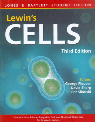
Lewin's CELLS, 3/e
Lewin's CELLS, 3/e
₹2,695.50 ₹2,995.00 Save: ₹299.50 (10%)
Go to cartISBN: 9789380853888
Bind: Paperback
Year: 2015
Pages: 1080
Size: 216 x 280 mm
Publisher: Jones & Bartlett Learning
Published in India by: Jones & Bartlett India
Exclusive Distributors: Viva Books
Sales Territory: India, Nepal, Pakistan, Bangladesh, Sri Lanka
Description:
The ideal text for undergraduate and graduate students in advanced cell biology courses
Extraordinary technological advances in the last century have fundamentally altered the way we ask questions about biology, and undergraduate and graduate students must have the necessary tools to investigate the world of the cell. The ideal text for students in advanced cell biology courses, Lewin's CELLS, Third Edition continues to offer a comprehensive, rigorous overview of the structure, organization, growth, regulation, movements, and interactions of cells, with an emphasis on eukaryotic cells. The text provides students with a solid grounding in the concepts and mechanisms underlying cell structure and function and will leave them with a firm foundation in cell biology as well as a "big picture" view of the world of the cell.
Revised and updated to reflect the most recent research in cell biology, Lewin's CELLS, Third Edition includes expanded chapters on Nuclear Structure and Transport, Chromatin and Chromosomes, Apoptosis, Principles of Cell Signaling, The Extracellular Matrix and Cell Adhesion, Plant Cell Biology, and more. All-new design features, a chapter-by-chapter emphasis on key concepts, and special feature boxes enhance pedagogy and emphasize retention and application of new skills. Thorough, accessible, and essential, Lewin's CELLS, Third Edition turns a new and sharper lens on the fundamental units of life.
Key features of the revised third edition include:
- Design features specifically intended to enhance pedagogy including Key Concepts, What's Next?, Concept and Reasoning Checks, and special feature boxes on Historical Perspectives, Medical Applications, and Methods and Techniques
- More student-friendly illustrations
Target Audience:
The book is designed for the students and academicians who are doing advanced Cell Biology courses.
Contents:
Feature Boxes • Preface • Acknowledgments • Contributors • Abbreviations
Part 1: Introduction
Chapter 1: What is a cell? (Vishwanath R. Lingappa and Benjamin Lewin) • 1.1 Introduction • 1.2 Life began as a self-replicating structure • 1.3 A prokaryotic cell consists of a single compartment • 1.4 Prokaryotes are adapted for growth under many diverse conditions • 1.5 A eukaryotic cell contains many membrane-delimited compartments • 1.6 Membranes allow the cytoplasm to maintain compartments with distinct environments • 1.7 The nucleus contains the genetic material and is surrounded by an envelope • 1.8 The plasma membrane allows a cell to maintain homeostasis • 1.9 Cells within cells: Organelles bounded by envelopes may have resulted from endosymbiosis • 1.10 DNA is the cellular hereditary material, but there are other forms of hereditary information • 1.11 Cells require mechanisms to repair damage to • DNA • 1.12 Mitochondria are energy factories • 1.13 Chloroplasts power plant cells • 1.14 Organelles require mechanisms for specific localization of proteins • 1.15 Proteins are transported to and through membranes • 1.16 Protein trafficking moves proteins through the endoplasmic reticulum and Golgi apparatus • 1.17 Protein folding and unfolding is an essential feature of all cells • 1.18 The shape of a eukaryotic cell is determined by its cytoskeleton • 1.19 Localization of cell structures is important • 1.20 Cellular functions are carried out by enzymes and enzyme pathways and are controlled by feedback mechanisms • 1.21 Signal transduction pathways execute predefined responses • 1.22 Many enzymes and signaling pathways are organized as multiprotein complexes that function as machines • 1.23 All organisms have cells that can grow and divide • 1.24 Differentiation creates specialized cell types, including terminally differentiated cells • References
Chapter 2: Bioenergetics and cellular metabolism (Bianca Barquera) • 2.1 Introduction • 2.2 Chemical equilibrium and reaction kinetics are linked • 2.3 The steady state model is essential for understanding the net flow of reactants in linked? reactions • 2.4 Thermodynamics is the systematic treatment of energy changes • 2.5 Standard free energy, the mass action ratio, and the equilibrium constant characterize reaction rates in metabolic pathways • 2.6 Glycolysis is the best understood metabolic pathway • 2.7 Pyruvate metabolism by the pyruvate dehydrogenase complex leads to oxidative respiration • Fatty acid oxidation is the major pathway of aerobic energy production • 2.9 The Krebs cycle oxidizes acetyl-CoA and is a metabolic hub • 2.10 Coupling of chemical reactions is a key feature of living organisms • 2.11 Oxidative phosphorylation is the final common pathway converting electron energy to ATP • 2.12 Photosynthesis completes the carbon cycle by converting CO2 to sugar • 2.13 Nitrogen metabolism encompasses amino acid, protein, and nucleic acid pathways • 2.14 The Cori cycle and the purine nucleotide cycle are specialized pathways • 2.15 Metabolic viewpoints provide insight into cellular regulation???only metabolically reversible reactions are possible regulatory sites • 2.16 Some new approaches • 2.17 Summary • References
Chapter 3: DNA replication, repair, and recombination (Jocelyn E. Krebs) • 3.1 Introduction • 3.2 DNA is the genetic material • 3.3 The structure of DNA • 3.4 DNA replication is semiconservative and bidirectional • 3.5 DNA polymerases replicate DNA • 3.6 Helicases, single-strand binding proteins, and topoisomerases are required for replication fork progression • 3.7 Priming is required to start DNA synthesis • 3.8 A sliding clamp ensures processive DNA replication • 3.9 Leading and lagging strand synthesis is coordinated • 3.10 Replication initiates at origins and is regulated by the cell cycle • 3.11 Replicating the ends of a linear chromosome • 3.12 DNA is subject to damage • 3.13 Direct repair can reverse some DNA damage • 3.14 Mismatch repair corrects replication errors • 3.15 Base excision repair replaces damaged bases • 3.16 Nucleotide excision repair removes bulky DNA lesions • 3.17 Double-strand breaks are repaired by homologous and nonhomologous pathways • 3.18 HR is used for both repair and meiotic recombination • 3.19 Summary • References
Chapter 4: Gene expression and regulation (David G. Bear) • 4.1 Introduction • 4.2 Genes are transcription units • 4.3 Transcription is a multistep process directed by DNA-dependent RNA polymerase • 4.4 RNA polymerases are large multisubunit protein complexes • 4.5 Promoters direct the initiation of transcription • 4.6 Activators and repressors regulate transcription initiation • 4.7 Transcriptional regulatory circuits control eukaryotic cell growth, proliferation, and differentiation • 4.8 The 5" and 3" ends of mature mRNAs are generated by RNA processing • 4.9 Terminators direct the end of transcription elongation • 4.10 Introns in eukaryotic pre-mRNAs are removed by the spliceosome • 4.11 Alternative splicing generates protein diversity • 4.12 Translation is a three-stage process that decodes an mRNA to synthesize a protein • 4.13 Translation is catalyzed by the ribosome • 4.14 Translation is guided by a large number of protein factors that regulate the interaction of aminoacylated tRNAs with the ribosome • 4.15 Translation is controlled by the interaction of the 5" and 3" ends of the mRNA and by translational repressor proteins • 4.16 Some mRNAs are translated at specific locations within the cytoplasm • 4.17 Sequence elements in the 5" and 3" untranslated regions determine the stability of an mRNA • 4.18 Noncoding RNAs are important regulators of gene expression • 4.19 What's next? • 4.20 Summary • References
Chapter 5: Protein structure and function (Stephen J. Smerdon) • 5.1 Introduction • 5.2 X-ray crystallography and structural biology • 5.3 Nuclear magnetic resonance • 5.4 Electron microscopy of biomolecules and their complexes • 5.5 Protein structure representations: a primer • 5.6 Proteins are linear chains of amino acids: primary structure • 5.7 Secondary structure: the fundamental unit of protein architecture • 5.8 Tertiary structure and the universe of protein folds • 5.9 Modular architecture and repeating motifs • 5.10 Quaternary structure and higher-order assemblies • 5.11 Enzymes are proteins that catalyze chemical reactions • 5.12 Posttranslational modifications and cofactors • 5.13 Dynamics, flexibility, and conformational changes • 5.14 Protein?protein and protein?nucleic acid interactions • 5.15 Function without structure? • 5.16 Structure and medicine • 5.17 What's next? Structural biology in the postgenomic era • 5.18 Summary • References
Part 2: Membranes and transport mechanisms
Chapter 6: Transport of ions and small molecules across membranes (Stephan E. Lehnart and Andrew R. Marks) • 6.1 Introduction • 6.2 Channels and carriers are the main types of membrane transport proteins • 6.3 Hydration of ions influences their flux through transmembrane pores • 6.4 Electrochemical gradients across the cell membrane generate the membrane potential • 6.5 K+ channels catalyze selective and rapid ion permeation • 6.6 Different K+ channels use a similar gate coupled to different activating or inactivating mechanisms • 6.7 Voltage-dependent Na+ channels are activated by membrane depolarization and translate electrical signals • 6.8 Epithelial Na+ channels regulate Na+ • homeostasis • 6.9 Plasma membrane Ca2+ channels activate intracellular and intercellular signaling processes • 6.10 Cl- channels serve diverse biologic functions • 6.11 Selective water transport occurs through aquaporin channels • 6.12 Action potentials are electrical signals that depend on several types of ion channels • 6.13 Cardiac and skeletal muscles are activated by excitation-contraction coupling • 6.14 Some glucose transporters are uniporters • 6.15 Symporters and antiporters mediate coupled transport • 6.16 The transmembrane Na+ gradient is essential for the function of many transporters 6.17 Some Na+ transporters regulate cytosolic or extracellular pH • 6.18 The Ca2+-ATPase pumps Ca2+ into intracellular storage compartments • 6.19 The Na+/K+-ATPase maintains the plasma membrane • Na+ and K+ gradients • 6.20 The F1F0-ATP synthase couples H+ movement to ATP • synthesis or hydrolysis • 6.21 H+-ATPases transport protons out of the cytosol • 6.22 What's next? • 6.23 Summary • 6.24 Supplement: Derivation and application of the Nernst equation • 6.25 Supplement: Most K+ channels undergo rectification • 6.26 Supplement: Mutations in an anion channel cause cystic fibrosis • References
Chapter 7: Membrane targeting of proteins (D. Thomas Rutkowski and Vishwanath R. Lingappa) • 7.1 Introduction • 7.2 Proteins enter the secretory pathway by translocation across the endoplasmic reticulum (ER) membrane (an overview) • 7.3 Proteins use signal sequences to target the ER for translocation • 7.4 Signal sequences are recognized by the signal recognition particle (SRP) • 7.5 Interaction between SRP and its receptor allows proteins to dock at the ER membrane • 7.6 The translocon is an aqueous channel that conducts proteins • 7.7 Translation is coupled to translocation for most eukaryotic secretory and transmembrane proteins • 7.8 Some proteins target and translocate posttranslationally • 7.9 ATP hydrolysis drives translocation • 7.10 Transmembrane proteins move out of the translocation channel and into the lipid bilayer • 7.11 Orientation of transmembrane proteins is determined as they are integrated into the membrane • 7.12 Signal sequences are removed by signal peptidase • 7.13 The lipid glycosylphosphatidylinositol (GPI) is added to some translocated proteins • 7.14 Sugars are added to many translocating proteins • 7.15 Chaperones assist folding of newly translocated proteins • 7.16 Protein disulfide isomerase ensures the formation of the correct disulfide bonds as proteins fold • 7.17 The calnexin/calreticulin chaperoning system recognizes carbohydrate modifications • 7.18 The assembly of proteins into complexes is monitored • 7.19 Terminally misfolded proteins in the ER are returned to the cytosol for degradation • 7.20 Communication between the ER and nucleus prevents the accumulation of unfolded proteins in the lumen 7.21 The ER synthesizes the major cellular phospholipids • 7.22 Lipids must be moved from the ER to the membranes of other organelles • 7.23 The two leaflets of a membrane often differ in lipid composition • 7.24 The ER is morphologically and functionally subdivided • 7.25 The ER is a dynamic organelle • 7.26 Signal sequences are also used to target proteins to other organelles • 7.27 Import into mitochondria begins with signal sequence recognition at the outer membrane • 7.28 Complexes in the inner and outer membranes cooperate in mitochondrial protein import • 7.29 Proteins imported into chloroplasts must also cross two membranes • 7.30 Proteins fold before they are imported into peroxisomes • 7.31 What's next? • 7.32 Summary • References
Chapter 8: Protein trafficking between membranes (Vivek Malhotra, Graham Warren, and Ira Mellman) • 8.1 Introduction • 8.2 Overview of the exocytic pathway • 8.3 Overview of the endocytic pathway • 8.4 Concepts in vesicle-mediated protein transport • 8.5 The concepts of signal-mediated and bulk flow protein transport • 8.6 Coat protein II (COPII)?coated vesicles mediate transport from the ER to the Golgi apparatus • 8.7 Resident proteins that escape from the ER are retrieved • 8.8 Coat protein I (COPI)?coated vesicles mediate • retrograde transport from the Golgi apparatus to the ER • 8.9 There are two popular models for forward transport through the Golgi apparatus • 8.10 Retention of proteins in the Golgi apparatus depends on the membrane-spanning domain • 8.11 Rab GTPases and tethers are two types of proteins that regulate vesicle targeting • 8.12 Soluble N-ethymaleimide-sensitive factor attachment protein receptor (SNARE) proteins likely mediate fusion of vesicles with target membranes • 8.13 Endocytosis is often mediated by clathrin-coated vesicles • 8.14 Adaptor complexes link clathrin and transmembrane cargo proteins • 8.15 Some receptors recycle from early endosomes, whereas others are degraded in lysosomes • 8.16 Early endosomes become late endosomes and lysosomes by maturation • 8.17 Sorting of lysosomal proteins occurs in the trans-Golgi network (TGN) • 8.18 Polarized epithelial cells transport proteins to apical and basolateral membranes • 8.19 Some cells store proteins for later secretion • 8.20 Some proteins are secreted without entering the ER?Golgi pathway • 8.21 What's next? • 8.22 Summary • References
Part 3: The nucleus
Chapter 9: Nuclear structure and transport (Charles N. Cole) • 9.1 Introduction • 9.2 Nuclei vary in appearance according to cell type and organism • 9.3 Chromosomes occupy distinct territories • 9.4 The nucleus contains subcompartments that are not membrane-bounded • 9.5 Some processes occur at distinct nuclear sites and may reflect an underlying structure • 9.6 The nucleus is bounded by the nuclear envelope • 9.7 The nuclear lamina underlies the nuclear envelope • 9.8 Large molecules are actively transported between the nucleus and cytoplasm • 9.9 Nuclear pore complexes are symmetrical channels • 9.10 Nuclear pore complexes are constructed from nucleoporins • 9.11 Proteins are selectively transported into the nucleus through nuclear pores • 9.12 Nuclear localization sequences target proteins to the nucleus • 9.13 Cytoplasmic receptors recognize nuclear localization sequences (NLSs) and mediate nuclear protein import • 9.14 Export of proteins from the nucleus is also receptor-mediated • 9.15 The Ran GTPase controls the directionality and irreversibility of nuclear transport • 9.16 Multiple models have been proposed for the mechanism of movement through nuclear pore complexes • 9.17 Nuclear transport can be regulated • 9.18 Multiple classes of RNA are exported from the nucleus • 9.19 Ribosomal subunits are assembled in the nucleolus and exported by multiple receptors • 9.20 tRNAs are exported by a dedicated exportin and can also use other receptors • 9.21 mRNAs are exported from the nucleus as RNA-protein complexes • 9.22 hnRNPs move from sites of processing to nuclear pore complexes • 9.23 mRNA export requires several novel factors • 9.24 U snRNAs are exported, modified, assembled into complexes, and imported back into the nucleus • 9.25 Precursors to microRNAs are partially processed in the nucleus, exported, and further processed in the cytoplasm • 9.26 What's next? • 9.27 Summary • References
Chapter 10: Chromatin and chromosomes (Benjamin Lewin and Jocelyn E. Krebs) • 10.1 Introduction • 10.2 Chromatin is divided into euchromatin and eterochromatin • 10.3 Chromosomes have banding patterns • 10.4 Eukaryotic DNA has loops and domains attached to a scaffold • 10.5 Specific sequences attach DNA to an interphase matrix or a metaphase scaffold • 10.6 The eukaryotic chromosome is a segregation device • 10.7 Point centromeres have short DNA sequences in Saccharomyces cerevisiae • 10.8 The centromere binds a protein complex • Regional centromeres contain repetitive DNA • 10.10 Telomeres are replicated by a special mechanism • 10.11 Lampbrush chromosomes are extended • 10.12 Polytene chromosomes form bands • 10.13 Polytene chromosomes expand at sites of gene expression • 10.14 The nucleosome is the subunit of all chromatin • 10.15 DNA is coiled in arrays of nucleosomes • 10.16 Nucleosomes have a common structure • 10.17 Organization of the histone octamer • 10.18 Histone variants produce alternative nucleosomes • 10.19 The path of nucleosomes in the chromatin fiber • 10.20 Replication of chromatin requires assembly of nucleosomes • 10.21 Do nucleosomes lie at specific positions? • 10.22 Domains of nuclease sensitivity define regions that contain active genes • 10.23 Histone octamers are displaced and reassembled during transcription • 10.24 Chromatin remodeling is an active process • 10.25 Histones are covalently modified • 10.26 Heterochromatin propagates from a nucleation event • 10.27 Heterochromatin depends on interactions with histones • 10.28 X chromosomes undergo global changes • 10.29 Chromosome condensation is caused by ondensins • 10.30 What's next? • 10.31 Summary • References
Part 4: The cytoskeleton
Chapter 11: Microtubules (Lynne Cassimeris) • 11.1 Introduction • 11.2 Microtubules are polar polymers of a- and ??-tubulin • 11.3 General functions of microtubules • 11.4 Purified tubulin subunits assemble into microtubules • 11.5 Microtubule assembly and disassembly proceed by a unique process termed dynamic instability • 11.6 A cap of GTP-tubulin subunits regulates the transitions of dynamic instability • 11.7 Cells use microtubule-organizing centers to nucleate microtubule assembly • 11.8 Microtubule dynamics in cells • 11.9 Why do cells have dynamic microtubules? • 11.10 Cells use several classes of proteins to regulate the stability of their microtubules • 11.11 Introduction to microtubule-based motor proteins • 11.12 How motor proteins work • 11.13 How cargoes are loaded onto the right motor • 11.14 Microtubule dynamics and motors combine to generate the asymmetric organization of cells • 11.15 Interactions between microtubules and actin filaments • 11.16 Cilia and flagella are motile structures • 11.17 What's next? • 11.18 Summary • 11.19 Supplement: What if tubulin did not hydrolyze GTP? • 11.20 Supplement: Fluorescence recovery after photobleaching • 11.21 Supplement: Tubulin synthesis and modification • References
Chapter 12: Actin (Enrique M. De La Cruz and E. Michael Ostap) • 12.1 Introduction • 12.2 Actin is a ubiquitously expressed cytoskeletal protein • 12.3 Actin monomers bind ATP and ADP • 12.4 Actin filaments are structurally polarized polymers • 12.5 Actin polymerization is a multistep and dynamic process • 12.6 Actin subunits hydrolyze ATP after polymerization • 12.7 Actin binding proteins regulate actin polymerization and organization • 12.8 Actin monomer binding proteins influence polymerization • 12.9 Nucleating proteins control cellular actin polymerization • 12.10 Capping proteins regulate the length of actin filaments • 12.11 Severing and depolymerizing proteins regulate actin filament dynamics • 12.12 Cross-linking proteins organize actin filaments into bundles and orthogonal networks • 12.13 Actin and actin binding proteins work together to drive cell migration • 12.14 Small G proteins regulate actin polymerization • 12.15 Myosins are actin-based molecular motors with essential roles in many cellular processes • 12.16 Myosins have three structural domains • 12.17 ATP hydrolysis by myosin is a multistep reaction • 12.18 Myosin motors have kinetic properties suited for their cellular roles • 12.19 Myosins take nanometer steps and generate piconewton forces • 12.20 Myosins are regulated by multiple mechanisms • 12.21 Myosin-II functions in muscle contraction • 12.22 What's next? • 12.23 Summary • References
Chapter 13: Intermediate filaments (Birgit Lane) • 13.1 Introduction • 13.2 Similarities in structure define the intermediate filament family • 13.3 Intermediate filament subunits assemble with high affinity into strain-resistant structures • 13.4 Two-thirds of all intermediate filament proteins are keratins • 13.5 Mutations in keratins cause epithelial cell fragility • 13.6 Intermediate filament proteins of nerve, muscle, and connective tissue often show overlapping expression • 13.7 Lamin intermediate filaments reinforce the nuclear envelope • 13.8 Even the divergent lens filament proteins are conserved in evolution • 13.9 Posttranslational modifications regulate and remodel intermediate filament networks • 13.10 Interacting proteins facilitate secondary functions of intermediate filaments • 13.11 Intermediate filament genes are represented through metazoan evolution • 13.12 What's next? • 13.13 Summary • References
Part 5: Cell division, apoptosis, and cancer
Chapter 14: Mitosis (Conly L. Rieder) • 14.1 Introduction • 14.2 Mitosis is divided into stages • 14.3 Mitosis requires the formation of a new apparatus called the spindle • 14.4 Spindle formation and function depend on the dynamic behavior of microtubules and their associated motor proteins • 14.5 Centrosomes are microtubule-organizing centers • 14.6 Centrosomes reproduce about the time the DNA is replicated • 14.7 Spindles begin to form as separating asters interact • 14.8 Spindles require chromosomes for stabilization but can "self-organize" without centrosomes • 14.9 The centromere is a specialized region on the chromosome that contains the kinetochores • 14.10 Kinetochores form at the onset of prometaphase and contain microtubule motor proteins5 • 14.11 Kinetochores capture and stabilize their associated microtubules • 14.12 Mistakes in kinetochore attachment are corrected • 14.13 Kinetochore fibers must both shorten and elongate to allow chromosomes to move • 14.14 Congression involves pulling forces that act on the kinetochores • 14.15 Congression is also regulated by forces that act along the chromosome arms and the activity of sister kinetochores • 14.16 Kinetochores control the metaphase-to-anaphase transition • The force to move a chromosome toward a pole is produced by two mechanisms • 14.18 Anaphase has two phases • 14.19 Changes occur during telophase that lead the cell out of the mitotic state • 14.20 During cytokinesis, the cytoplasm is partitioned to form two new daughter cells • 14.21 Formation of the contractile ring requires the spindle and stem bodies • 14.22 The contractile ring cleaves the cell in two • 14.23 The segregation of nonnuclear organelles during cytokinesis is based on chance • 14.24 What's next? • 14.25 Summary • References
Chapter 15: Cell cycle regulation (Kathleen L. Gould and Susan L. Forsburg) • 15.1 Introduction • 15.2 Several experimental systems are used for cell cycle research • 15.3 A cycle of cyclin-dependent kinase activities regulates cell proliferation • 15.4 CDK-cyclin complexes are regulated in several ways • 15.5 Cells may withdraw from the cell cycle • 15.6 Entry into cell cycle is tightly regulated • 15.7 DNA replication requires the ordered assembly of protein complexes • 15.8 Mitosis is orchestrated by several protein kinases • 15.9 Sister chromatids are held together until anaphase • 15.10 Exit from mitosis requires more than cyclin proteolysis • 15.11 Events of the cell cycle are coordinated • 15.12 Checkpoint controls coordinate different cell cycle events • 15.13 DNA replication and DNA damage checkpoints monitor defects in DNA metabolism • 15.14 The SAC monitors defects in chromosomemicrotubule attachment • 15.15 Cell cycle deregulation can lead to cancer • 15.16 What's next? • 15.17 Summary • References
Chapter 16: Apoptosis (Douglas R. Green) • 16.1 Introduction • 16.2 Caspases orchestrate apoptosis by cleaving specific substrates • 16.3 Executioner caspases are activated by cleavage, whereas initiator caspases are activated by dimerization • 16.4 Some inhibitors of apoptosis proteins block caspases • 16.5 Some caspases have functions in inflammation • 16.6 The death receptor pathway of apoptosis transmits external signals • 16.7 Apoptosis signaling by TNFR1 is complex • 16.8 Mitochondrial pathway of apoptosis • 16.9 Bcl-2 family proteins mediate and regulate MOMP and apoptosis • 16.10 Multidomain Bcl-2 proteins Bax and Bak are required for MOMP • 16.11 Activation of Bax and Bak are controlled by other Bcl-2 family proteins • 16.12 Cytochrome c, released upon MOMP, induces caspase activation • 16.13 Some proteins released upon MOMP block IAPs • 16.14 The death receptor pathway of apoptosis can engage MOMP through the cleavage of the BH3-only protein Bid • 16.15 MOMP can cause caspase-independent cell death • 16.16 Mitochondrial permeability transition can cause MOMP • 16.17 Many discoveries about apoptosis were made in nematodes • 16.18 Apoptosis in insects has features distinct from mammals and nematodes • 16.19 Clearance of apoptotic cells requires cellular interaction • 16.20 Apoptosis plays a role in diseases such as viral infection and cancer • 16.21 Apoptotic cells are gone but not forgotten • 16.22 What's next? • 16.23 Summary • References
Chapter 17: Cancer: Principles and overview (Robert A. Weinberg) • 17.1 Tumors are masses of cells derived from a single cell • 17.2 Cancer cells have a number of phenotypic characteristics • 17.3 Cancer cells arise after DNA damage • 17.4 Cancer cells are created when certain genes are mutated • 17.5 Cellular genomes harbor a number of proto-oncogenes • 17.6 Elimination of tumor-suppressor activity requires two mutations • 17.7 Genesis of tumors is a complex process • 17.8 Cell growth and proliferation are activated by growth factors • 17.9 Cells are subject to growth inhibition and may exit from the cell cycle • 17.10 Tumor suppressors block inappropriate entry into the cell cycle • 17.11 Mutation of DNA repair and maintenance genes can increase the overall mutation rate • 17.12 Cancer cells may achieve immortality • 17.13 Access to vital supplies is provided by angiogenesis • 17.14 Cancer cells may invade new locations in the body • 17.15 What's next? • 17.16 Summary • References
Part 6: Cell communication
Chapter 18: Principles of cell signaling (Elliott M. Ross and Melanie H. Cobb) • 18.1 Introduction • 18.2 Cellular signaling is primarily chemical • 18.3 Receptors sense diverse stimuli but initiate a limited repertoire of cellular signals • 18.4 Receptors are catalysts and amplifiers • 18.5 Ligand binding changes receptor conformation • 18.6 Signals are sorted and integrated in signaling pathways and networks • 18.7 Cellular signaling pathways can be thought of as biochemical logic circuits • 18.8 Scaffolds increase signaling efficiency and enhance spatial organization of signaling • 18.9 Independent, modular domains specify protein-protein interactions • 18.10 Cellular signaling is remarkably adaptive • 18.11 Signaling proteins are frequently expressed as multiple species • 18.12 Activating and deactivating reactions are separate and independently controlled • 18.13 Cellular signaling uses both allostery and covalent modification • 18.14 Second messengers provide readily diffusible pathways for information transfer • 18.15 Ca2+ signaling serves diverse purposes in all eukaryotic cells • 18.16 Lipids and lipid-derived compounds are signaling molecules • 18.17 PI3-kinase regulates both cell shape and the activation of essential growth and metabolic functions • 18.18 Signaling through ion channel receptors is very fast • 18.19 Nuclear receptors regulate transcription • 18.20 G protein-signaling modules are widely used and highly adaptable • 18.21 Heterotrimeric G proteins regulate a wide variety of effectors • 18.22 Heterotrimeric G proteins are controlled by a regulatory GTPase cycle • 18.23 Small, monomeric GTP-binding proteins are multiuse switches • 18.24 Protein phosphorylation/dephosphorylation is a major regulatory mechanism in the cell • 18.25 Two-component protein phosphorylation systems are signaling relays • 18.26 Pharmacologic inhibitors of protein kinases may be used to understand and treat disease • 18.27 Phosphoprotein phosphatases reverse the actions of kinases and are independently regulated • 18.28 Covalent modification by ubiquitin and ubiquitin-like proteins is another way of regulating protein function • 18.29 Wnt pathway regulates cell fate during development and other processes in the adult • 18.30 Diverse signaling mechanisms are regulated by protein-tyrosine kinases • 18.31 Src family protein kinases cooperate with receptor protein-tyrosine kinases • 18.32 MAPKs are central to many signaling pathways • 18.33 Cyclin-dependent protein kinases control the cell cycle • 18.34 Diverse receptors recruit protein-tyrosine kinases to the plasma membrane • 18.35 What's next? • 18.36 Summary • References
Chapter 19: The extracellular matrix and cell adhesion (George Plopper) • 19.1 Introduction • 19.2 Collagen provides structural support to tissues • 19.3 Fibronectins connect cells to collagenous matrices • 19.4 Elastic fibers impart flexibility to tissues • 19.5 Laminins provide an adhesive substrate for cells • 19.6 Matricellular proteins regulate cell?ECM interactions • 19.7 Proteoglycans provide hydration to tissues • 19.8 HA is a GAG enriched in connective tissues • 19.9 HSPGs are cell surface coreceptors • 19.10 Basal lamina is a specialized ECM5 • 19.11 Proteases degrade ECM components • 19.12 Most integrins are receptors for ECM proteins • 19.13 Integrin receptors participate in cell signaling • 19.14 Integrins and ECM molecules play key roles in development • 19.15 Tight junctions form selectively permeable barriers between cells • 19.16 Septate junctions in invertebrates are similar to tight junctions • 19.17 Adherens junctions link adjacent cells • 19.18 Desmosomes are intermediate filament-based cell adhesion complexes • 19.19 Hemidesmosomes attach epithelial cells to the basal lamina • 19.20 Gap junctions allow direct transfer of molecules between adjacent cells • 19.21 Calcium-dependent cadherins mediate adhesion between cells • 19.22 Calcium-independent neural cell adhesion molecules mediate adhesion between neural cells • 19.23 Selectins control adhesion of circulating immune cells • 19.24 What's next? • 19.25 Summary • References
Part 7: Prokaryotic and plant cells
Chapter 20: Prokaryotic cell biology (Matthew Chapman and Jeff Errington) • 20.1 Introduction • 20.2 Molecular phylogeny techniques are used to understand microbial evolution • 20.3 Prokaryotic life-styles are diverse • 20.4 Archaea are prokaryotes with similarities to eukaryotic cells • 20.5 Most prokaryotes produce a polysaccharide-rich layer called the capsule • 20.6 The bacterial cell wall contains a cross-linked meshwork of peptidoglycan • 20.7 The cell envelope of gram-positive bacteria has unique features • 20.8 Gram-negative bacteria have an outer membrane and a periplasmic space • 20.9 The cytoplasmic membrane is a selective barrier for secretion • 20.10 Prokaryotes have several secretion pathways • 20.11 Pili and flagella are appendages on the cell surface of most prokaryotes • 20.12 Prokaryotic genomes contain chromosomes and mobile DNA elements • 20.13 The bacterial nucleoid and cytoplasm are highly ordered • 20.14 Bacterial chromosomes are replicated in specialized replication factories • 20.15 Prokaryotic chromosome segregation occurs in the absence of a mitotic spindle • 20.16 Prokaryotic cell division involves formation of a complex cytokinetic ring • 20.17 Prokaryotes respond to stress with complex developmental changes • 20.18 Some prokaryotic life cycles include obligatory developmental changes • 20.19 Some prokaryotes and eukaryotes have endosymbiotic relationships • 20.20 Prokaryotes can colonize and cause disease in higher organisms • 20.21 Biofilms are highly organized communities of microbes • 20.22 What's next? • 20.23 Summary • References
Chapter 21: Plant cell biology (Clive Lloyd) • 21.1 Introduction • 21.2 How plants grow • 21.3 The meristem provides new growth modules in a repetitive manner • 21.4 The plane in which a cell divides is important for tissue organization • 21.5 Cytoplasmic structures predict the plane of cell division before mitosis begins • 21.6 Plant mitosis occurs without centrosomes • 21.7 The cytokinetic apparatus builds a new wall in the plane anticipated by the preprophase band • 21.8 Secretion during cytokinesis forms the cell plate • 21.9 Plasmodesmata are intercellular channels that connect plant cells • 21.10 Cell expansion is driven by swelling of the vacuole • 21.11 The large forces of turgor pressure are resisted by the strength of cellulose microfibrils in the cell wall • 21.12 The cell wall must be loosened and reorganized to allow growth • 21.13 Cellulose is synthesized at the plasma membrane, not preassembled and secreted like other wall components • 21.14 Cortical microtubules organize components in the cell wall • 21.15 Cortical microtubules are highly dynamic and can change their orientation • 21.16 A dispersed Golgi system delivers vesicles to the cell surface for growth • 21.17 Actin filaments form a network for delivering materials around the cell • 21.18 Differentiation of xylem cells requires extensive specialization • 21.19 Specialized plant cells elongate by tip growth • 21.20 Plants contain unique organelles called plastids • 21.21 Chloroplasts manufacture food from atmospheric CO2 • 21.22 Polarized flow of the hormone auxin coordinates growth • 21.23 What's next? • 21.24 Summary • References
Glossary
Index
About the Authors:
George Plopper, PhD-Rensselaer Polytechnic Institute
George Plopper is a Professor in the Department of Biology at Rensselaer Polytechnic Institute in Troy, New York. He received his Bachelor of Arts in General Biology from the University of California, San Diego. He completed his PhD in Cell & Developmental Biology at Harvard University, then completed his postdoctoral training in Cell Biology at The Scripps Research Institute in La Jolla, California. Dr. Plopper served as Assistant Professor at The University of Nevada, Las Vegas before moving to his present position at Rensselaer Polytechnic Institute. He has taught cell biology to undergraduate and graduate students since 1985, receiving four teaching awards. He was named a National Academies Education Fellow in the Life Sciences by the National Academy of Sciences in 2004.
David Sharp, PhD-Professor of Physiology & Biophysics, Albert Einstein College of Medicine
Eric Sikorski, PhD-University of Tampa
